Bones In Your Leg Diagram - Bone structure of the lower leg.. This keeps the bones together, giving a high ankle sprain time to heal. This can cause tenderness in the right or left buttock and severe pain down the leg. The proximal portion of the tibia is tibial plateau which acts as a cusp for the knee, the distal portion tapers into the medial malleoli and the concave surface which articulates with the talus at the ankle joint. There are a total of 60 bones in the legs. In the leg muscles diagram above, there are many muscles that make up your legs and support it to move.
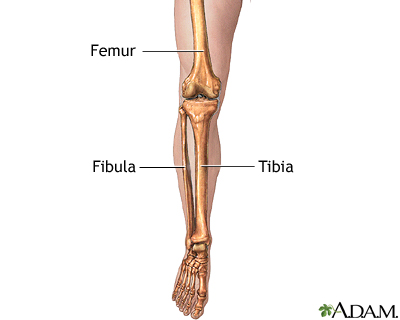
The lower leg extends from the knee to the ankle. The fibula, or calf bone, is smaller and is located on the outside of the lower leg. Distal to the ankle is the foot. Its lower end helps create the knee joint. The femur, or thighbone, is the longest and largest bone in the human body.
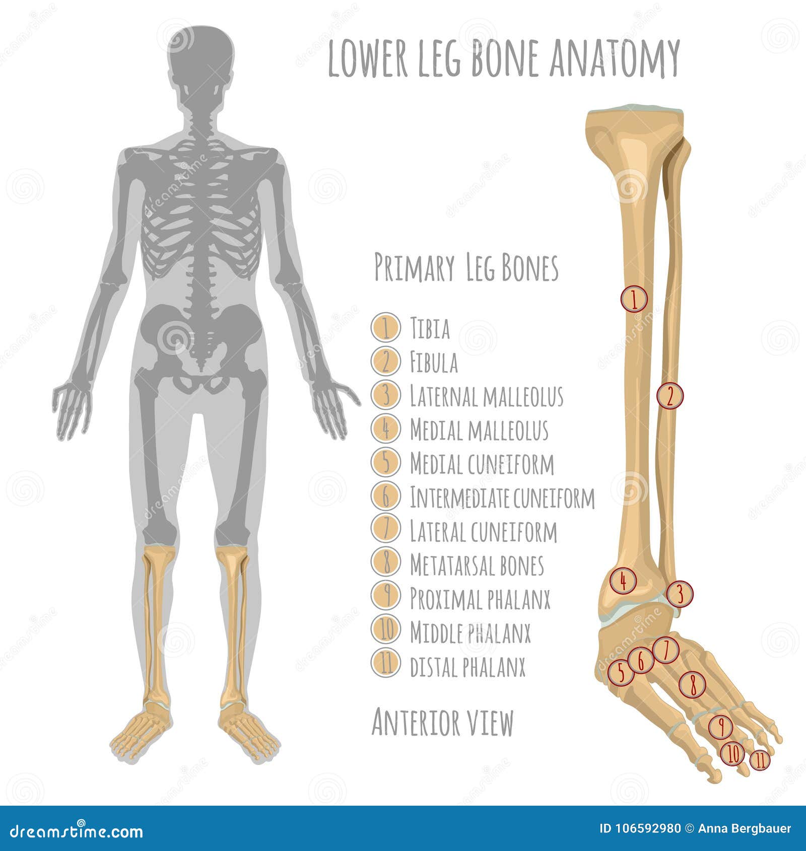
Leg Skeletal Anatomy Medlineplus Medical Encyclopedia Image from medlineplus.gov In addition, the broad hip bones provide protection to the delicate internal organs of the pelvis, such as the intestines, urinary bladder, and uterus. The second metatarsal bone is the longest. The lower leg is comprised of two bones, the tibia and the smaller fibula. The femur is the single bone of the thigh. The tibia (also called the shinbone) is located near the midline of the leg. The knee joint is the largest joint in the body and is primarily a hinge joint, although some sliding and rotation occur. The pelvic region is the area between the trunk — or main body — and the lower extremities, or legs. Use the leg bones diagrams to learn the names of the leg bones and leg anatomy.
The male pelvis is different from a female's.
Fpe medical review board a foot pain diagram is a great tool to help you work out what is causing your ankle and foot pain. The sacrum and the coccyx attach to the two hip bones to form the pelvis, but are more important to the spinal column, where they are counted. The bones of the leg are the femur, tibia, fibula and patella.the foot bones shown in this diagram are the talus, navicular, cuneiform, cuboid, metatarsals and calcaneus. Your hamstring is directly connected to your ischium bones, and any tear or damage to your hamstring can result in sit bone pain. The male pelvis is different from a female's. The pelvic region is the area between the trunk — or main body — and the lower extremities, or legs. This allows weight to be distributed either anteriorly or posteriorly throughout the foot. Bones, muscles, tendons and nerves which will each give slightly different foot pain symptoms. Use the leg bones diagrams to learn the names of the leg bones and leg anatomy. This can cause tenderness in the right or left buttock and severe pain down the leg. The majority of muscles in the leg are considered long muscles, in that they stretch great distances. Its lower end helps create the knee joint. The second metatarsal bone is the longest.
The first metatarsal bone, the shortest, thickest and strongest metatarsal, links to the big toe. Chloe wilson bsc(hons) physiotherapy reviewed by: Fpe medical review board a foot pain diagram is a great tool to help you work out what is causing your ankle and foot pain. Diagram depicting the arterial supply to a growing leg. Your hamstring is directly connected to your ischium bones, and any tear or damage to your hamstring can result in sit bone pain.
Pin By Genna Hornsby On Anatomy Medical Anatomy Anatomy Bones Human Anatomy And Physiology from i.pinimg.com These are the femur, patella, tibia, fibula, tarsal bones, metatarsal bones, and phalanges (see figure 6.51). The smaller bone that runs alongside the tibia (fibula) and the kneecap (patella) are the other bones that make the knee joint. The talocrual joint is made up of three main bones. It is located toward the middle of the lower leg. The medial, larger bone of the lower leg. The tibia and fibula are two long bones that run parallel to each other, forming the scaffold of the leg and providing attachment points for many muscles. Let's review all of these bones one last time. The lower limb contains 30 bones.
Bone structure of the lower leg.
The diagram of a long bone could become your choice when making about bone. Fpe medical review board a foot pain diagram is a great tool to help you work out what is causing your ankle and foot pain. Ankle & lower leg anatomy. The tibia and fibula are two long bones that run parallel to each other, forming the scaffold of the leg and providing attachment points for many muscles. The pelvic bones are smaller and. The smaller bone that runs alongside the tibia (fibula) and the kneecap (patella) are the other bones that make the knee joint. The first metatarsal bone, the shortest, thickest and strongest metatarsal, links to the big toe. Treatment of a broken leg depends on the location and severity of the injury. This allows weight to be distributed either anteriorly or posteriorly throughout the foot. The lower leg contains two major long bones, the tibia and the fibula, which are both very strong skeletal structures. The largest and most medial leg bone, forming both the knee and ankle joints. The lower leg extends from the knee to the ankle. Tendons connect the knee bones to the leg muscles that move the knee.
The second metatarsal bone is the longest. This area is commonly referred to as the calf. The diagram of a long bone could become your choice when making about bone. Cat leg bone diagram : The femur is the single bone of the thigh.
Anatomy Lower Leg Bones Stock Illustrations 392 Anatomy Lower Leg Bones Stock Illustrations Vectors Clipart Dreamstime from thumbs.dreamstime.com Ankle & lower leg anatomy. The pelvic bones are smaller and. The bones together make up the hip. The tibia and fibula are two long bones that run parallel to each other, forming the scaffold of the leg and providing attachment points for many muscles. The lower leg extends from the knee to the ankle. Fpe medical review board a foot pain diagram is a great tool to help you work out what is causing your ankle and foot pain. The lower leg lies between the knee and the ankle. The hip itself is a ball and socket joint, much like the shoulder.the structures necessary to create this joint are the socket, the joint capsule, muscle, ligaments, and the neck.
Fpe medical review board a foot pain diagram is a great tool to help you work out what is causing your ankle and foot pain.
The fibula, or calf bone, is smaller and is located on the outside of the lower leg. The human leg consists of 8 bones, 4 per leg. The bones of the leg and foot form part of the appendicular skeleton that supports the many muscles of the lower limbs. Almost every skeletal muscle works by pulling two or more bones either closer together or further apart. Bone structure of the lower leg. The hip itself is a ball and socket joint, much like the shoulder.the structures necessary to create this joint are the socket, the joint capsule, muscle, ligaments, and the neck. Treatment of a broken leg depends on the location and severity of the injury. Cat leg bone diagram : The largest and most medial leg bone, forming both the knee and ankle joints. Your hamstring is directly connected to your ischium bones, and any tear or damage to your hamstring can result in sit bone pain. This keeps the bones together, giving a high ankle sprain time to heal. The talocrual joint is made up of three main bones. The male pelvis is different from a female's.
The largest and most medial leg bone, forming both the knee and ankle joints leg bones diagram. These are the femur, patella, tibia, fibula, tarsal bones, metatarsal bones, and phalanges (see figure 6.51).


Posting Komentar
0 Komentar